
Giant fibroepithelial breast polyp
DOI:
https://doi.org/10.51496/jogm.v4.S1.193Keywords:
fibroepithelial polyps, large, surgery, acrochordonsAbstract
Introduction: Fibroepithelial polyps are common benign skin lesions
rarely noted in the female nipple. In this location, they have the potential
to ulcerate and cause bloody discharge.
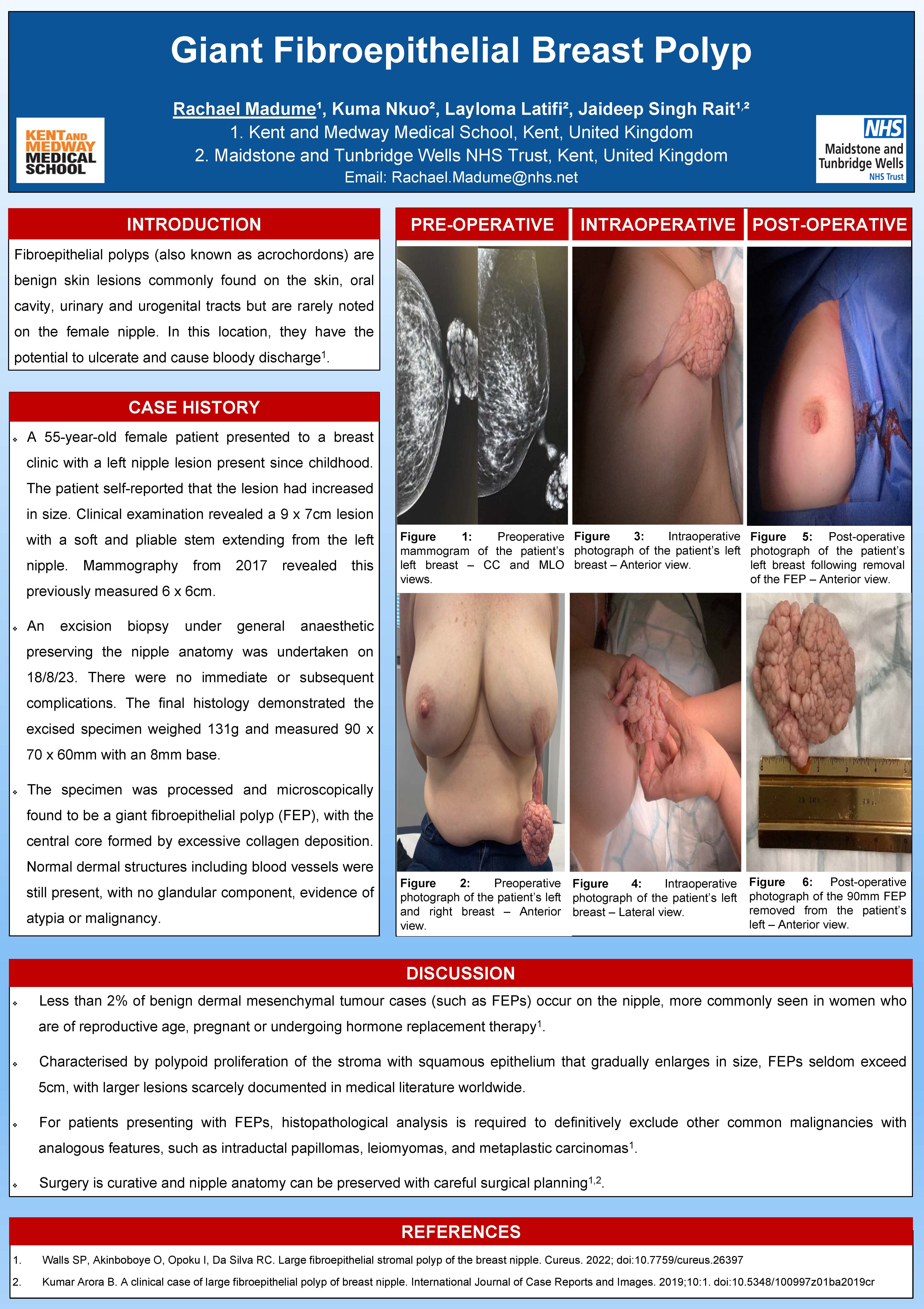
Case history: A 55-year-old female patient presented to a breast clinic
with a left nipple lesion present since childhood. The patient self-reported
that the lesion had increased in size. Clinical examination revealed a 9 ×
7 cm lesion with a soft and pliable stem extending from the left nipple.
Mammography from 2017 revealed this previously measured 6 × 6 cm.
An excision biopsy under general anaesthetic preserving the nipple
anatomy was undertaken on 18/8/23. There were no immediate or subsequent
complications. The final histology demonstrated the excised specimen
weighed 131 g and measured 90 × 70 × 60 mm with an 8 mm base.
The specimen was processed and microscopically found to be a giant
fibroepithelial polyp (FEP). The central core of the lesion was formed by
excessive collagen deposition. Normal dermal structures including blood
vessels were still present with no glandular component and there was no
evidence of atypia or malignancy.
Discussion: FEPs are benign dermal mesenchymal tumours also known
as acrochordons. They are noted most commonly on the neck, axilla,
perineum, and thighs but have also been found in the vagina, vulva, or
cervix and present in < 2% of cases in the nipple area.
FEPs rarely grow larger than 5 cm, with larger lesions rarely reported
in medical literature worldwide. It is possible to excise these lesions and
preserve the nipple anatomy with careful surgical planning.
Published
How to Cite
Issue
Section
Categories
License
Copyright (c) 2024 Rachael Madume, Kuma Nkuo, Layloma Latifi, Jaideep Singh Rait

This work is licensed under a Creative Commons Attribution 4.0 International License.


 Journal of Global Medicine | Editor-in-Chief: Olufunso Adedeji. MBBS, MD, FRCSEd.